
We recommend yearly screening and parasite prevention from approximately April-Nov.
(Ticks can be active in any weather above 4 degrees!)
If you visit areas with a warmer climate during the winter months we recommend continuing prevention all year.
We have many options of parasite prevention available to fit your pet's individual lifestyle, please call and ask us which one would best suit your pet's needs.

Positive cases reported in 2017
https://www.petdiseasereport.com/
Heartworm: The Parasite
By Wendy C. Brooks DVM, DABVP c/o www.veterinarypartner.com
Heartworm is a parasite that most dog owners and many cat owners have to be concerned about. The more you know, the better protected your pet can become. We have put together an information center to take you through the parasite's biology, the preventive medications, diagnosis, and treatment.
What is a Heartworm?
Heartworm (Dirofilaria immitis) is a fairly large worm up to 14 inches long that, in adulthood, lives in the heart and pulmonary arteries of an infected dog. Dogs acquire this infection through mosquito bites as mosquitoes readily pick up larval heartworms from infected dogs and carry them to new dogs. Some geographic areas have severe heartworm problems while other areas have none. In order for the parasite to establish its presence in an area, the following conditions must be met:
- Types of mosquitoes capable of carrying larval heartworms must be present
- The weather must be warm enough to allow heartworm larval development within the mosquito
- There must be infected dogs (or coyotes) in the area
- There must be vulnerable host dogs in the area
When these conditions come together, an area becomes endemic for heartworm disease.
The Detailed Version of the Heartworm Story: Let's Follow the Worm's Life Cycle
The Adult Heartworm
The adult heartworm is fairly large, several inches in length, and it prefers to live, not in the heart, but in the pulmonary arteries. It swims into a cozy tubular artery, where it is massaged and nourished by the blood coursing past it. In the pulmonary arteries of an infected dog, the worm's presence generates a strong inflammatory response and a tendency for blood to inappropriately clot. If enough worms are present, the heart must work extra hard to pump blood through the plugged up arteries.
If the worm infection is a heavy one (over 25 worms for a 40 lb. dog), the worms begin to back up into the right ventricle (the chamber that pumps blood through the lung). The worms actually take up a significant amount of space within the heart, leading to less blood being pumped.
When over 50 worms are present, the ventricle is full and the atrium, the chamber receiving blood from the rest of the body begins to contain worms.
When over 100 worms are present, the entire right side of the heart is filled with worms and there is very little room for any blood to be pumped. This drastic phenomenon is called Caval Syndrome and most dogs do not survive it.
Microfilariae (First Stage Larvae)
With adult male and female worms present, mating begins to occur. Heartworms do not lay eggs like other worm parasites; instead they give live birth and the baby worms are called Microfilariae. Microfilariae are released into the circulatory system in hope that they will be slurped up by a mosquito taking a blood meal and carried to a new host. Microfilariae may live up to 2 years within the host dog in whom they were born; if after this period a mosquito has not picked them up, they die of old age. Microfilariae may also be transmitted across the placental barrier to unborn puppies if the mother dog is infected with heartworm. It is important to realize that such puppies will not develop adult heartworms or heartworm disease from these microfilariae; in order for a heartworm to reach adulthood, it must be passed through a mosquito.
Parasitic worms have 5 larval stages and are termed L1, L2, L3, etc. Heartworm microfilariae are first stage larvae: L1s.
Note: Heartgard30 and Interceptor, the main heartworm preventives available commercially, will kill microfilariae. Dogs on heartworm preventive, even if infected with adult heartworms, will not test positive for microfilariae.
Inside the Mosquito
Within the mosquito's body, the microfilariae will develop to L2s and finally to L3s, the stage capable of infecting a new dog. How long this takes depends on the environmental conditions. In general, it takes a few weeks. A minimum environmental temperature of 57 degrees F is required throughout this period. The process goes faster in warmer weather.
Infecting a New Dog
When a dog is bitten by an infected mosquito, the L3 is not deposited directly into the dog's bloodstream. Instead, it is deposited in a tiny drop of mosquito "spit" adjacent to the mosquito bite. For transmission to occur, there must be adequate humidity to prevent evaporation of this fluid droplet before the L3s can swim through the mosquito bite and into the new host.
Once safely inside the new host, the L3 will spend the next week or two developing into an L4 within the host's skin.
The L4 will live in the skin for 3 months or so until it develops to the L5 stage and is ready to enter the host's circulatory system. The L5, which is actually a young adult, migrates to the heart and out into the pulmonary arteries (if there is room) where it will mate, approximately 5 to 7 months after first entering the new host.
Note: L3s are readily killed by Interceptor but not by Heartgard30. Interceptor and Heartgard30 both act primarily on the L4s living in the skin. After a dose of either medication, any L4s present will be wiped out. Heartgard30 is also able to kill the younger L5s.
What Happens in Heartworm Disease
Heartworm Disease vs. Heartworm Infection
Before reviewing the clinical signs seen in heartworm disease, an important distinction must be made between heartworm disease and heartworm infection. Heartworm infection by definition means the host animal (generally a dog) is parasitized by at least one life stage of the heartworm (Dirofilaria immitis). Dogs with heartworms in their bodies do not necessarily have adult worms in their hearts; they may have larval heartworms in their skin only. Dogs with heartworms in their bodies are not necessarily sick, either. Dogs with only larvae of one stage or another are not sick and it is controversial how dangerous it is for a dog to have only one or two adult heartworms. These dogs are certainly infected but they do not have heartworm disease.
On the other hand, dogs with heartworm disease are sick. They not only have the infection but they have any of the problems listed below because of it. Fortunately, heartworm disease is both treatable and preventable. Further sections of this web area explain both treatment and prevention; we will now discuss the damage heartworms can do to a dog's body.
Damage to the Pulmonary Arteries
Arteries do not do well having worms living inside them. The lining of the artery becomes damaged within days of the worm's arrival. Cells of the immune system are called into the area but the worm is far too big for these tiny cells to destroy. The resulting inflammation; however, continues to damage the artery. The arteries dilate and become tortuous (which may be visible on a radiograph). Aneurysms and abnormal blood clotting (embolism) results. Blood is shunted to other arteries which are not plugged up by worms and fluid begins to accumulate in the lung around the worm-filled arteries. Blood being sent to the lung is not efficiently oxygenated and areas of lung become consolidated and unable to participate in providing oxygen to the blood.
- Coughing and exercise intolerance result as areas of the lung are lost to the blood oxygenation process.
- Nose bleeds may occur due to abnormal blood clotting in the lung.
- A form of non-infectious pneumonia ("pulmonary eosinophilic granulomatosis") can result from excessive infiltration of inflammatory cells into the lung in response to the parasite.
Heart Failure
Blood normally is pumped with ease through the arteries of the lung. With the arteries plugged with worms, the heart must pump harder against the pressure of the plugged arteries. This condition is called "pulmonary hypertension" and the right side of the heart must drastically increase its ability to work. It may be strong enough and it may not.
If worms begin backing up into the heart, there will be less space in the pumping chamber for blood to be pumped. The heart must pump through the high pressure system of the plugged arteries using less blood then normal. In order to meet the body's oxygen demand, the heart must pump faster and stronger still. There may come a point when the heart simply is not strong enough.
- When the heart muscle begins to thicken (as any over-worked muscle will), it may not conduct electrical impulses normally. This means that the pumping/filling rhythm can be disrupted and an "ARRHYTHMIA" may result. IN ANY HEART DISEASE, ARRHYTHMIA IS A POSSIBILITY; WHEN ARRHYTHMIA IS A POSSIBILITY, SO IS SUDDEN DEATH.
- If the right side of the heart becomes too weak to keep up, fluid may accumulate in the chest cavity and abdominal cavity, leading to a pot-bellied appearance and/or difficulty breathing.
Development of Heartworm Disease
Chronic Immune Stimulation
When a dog goes without treatment for heartworm disease, its immune system becomes chronically stimulated. Antibodies, which are not only important tools of the immune system but are inflammatory proteins, are produced in high amounts all the time. These antibodies can cause a lot of trouble by precipitating in the delicate membranes of the eye, kidney, blood vessels, and joints. Antibodies stuck in these areas, call in inflammatory cells and damage these delicate membranes thus setting up tremendous tissue damage and pain.
Caval Syndrome
Caval syndrome represents an especially disastrous form of heartworm disease. Here, there are so many worms present (around 100) that the entire right side of the heart is filled with worms and they are backing out into the large veins that feed the right side of the heart. Usually there have been no signs of heart disease prior to the collapse, shock, and red blood cell destruction associated with this syndrome. Death usually occurs within 1 to 2 days and the only effective treatment is to open the dog's jugular vein and physically remove the worms with a special clamp. If enough worms can be removed to re-establish blood flow, the dog may survive.
Heartworm disease is a highly significant problem and must be managed both by dealing with the worms and by dealing with the heart disease.
Ticks and Lyme Disease
By Wendy C. Brooks, DVM, DipABVP
Educational Director, VeterinaryPartner.com
While the infection we know today as Lyme disease (named for the Connecticut town of Lyme) has been around for at least a century, public awareness and confusion did not really occur until the late 1980s. Media exposure of this infection virtually exploded, leaving most of the general public with some basic knowledge and concern about this infection as it pertains to themselves as well as to their dogs. The canine experience of Lyme disease is very different from the human infection and we hope to sort out confusion here.
Human Lyme Disease vs. Canine Lyme Disease
The Erythema migrans is the characteristic skin rash shown by humans shortly after Borrelia burgdorferi infection. Dogs only rarely exhibit a similar rash with Lyme disease. Photo by CDC.
The first lesson to be learned about the Lyme disease infection is that, as mentioned, it manifests completely differently in man’s best friend compared with the human experience. After being bitten by a tick that has transmitted Borrelia burgdorferi, 80 percent of humans will develop a rash and/or flu-like symptoms. In the next few weeks, joint pain ensues with 15 percent of people developing neurologic abnormalities associated with Lyme disease and five percent of people developing a heart rhythm disturbance called A-V block. At this same point in the infection timeline, dogs have yet to develop any symptoms at all and 90 percent of infected dogs never will.
Weeks to months after infection, about 60 percent of people will experience intermittent arthritis attacks and five percent will develop chronic neurologic manifestations. In humans, Lyme disease has the potential for serious long-term illness.
When canine illness does occur, it does not begin to manifest for weeks to months after infection at which point arthritis signs are noticed. Sometimes there is a fever. In dogs, heart and neurologic issues are exceedingly rare, plus the symptoms of canine Lyme disease generally respond rapidly to an inexpensive course of proper antibiotics (see below for details).
The Borrelia burgdorferi organism is fairly well suited to live in the canine body without causing trouble. Most exposed dogs harbor the organism uneventfully. Still, it is important not to discount Lyme disease in dogs completely lest you overlook an easily eliminated cause of chronic joint disease, especially in dogs of the Northeast U.S.
A dog’s most serious long-term potential regards glomerular disease. This is a type of kidney damage that occurs when the immune system is stimulated over a long time by a latent infectious organism or other immune stimulus. This is a much more insidious problem for which specific testing is needed (see below).
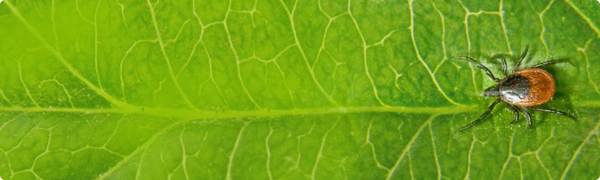
The Tick and Its Control
An organism that serves to transport and deliver an infectious organism from one host to another is called a vector. The vector of Lyme disease in the Northeastern United States is the deer tick, Ixodes scapularis. The female tick lays a clump of approximately 2,000 eggs in the spring. A small six-legged larva hatches and attaches to a host as soon as it is able. Since the larva is small, it typically can only reach a small host, usually a white-footed mouse. If the mouse is carrying the Lyme disease spirochete, the larva can get infected at this point.
Three stages of the deer tick: The larva (bottom right), nymph (bottom left), and adult female (top). Photo by CDC
When the larva is full of blood, it will drop off the host and lie dormant until the following spring, about a year later. At this point the larva molts and becomes a nymph. The nymph is a bit larger and may select another mouse as host or may approach larger game such as a dog or human. The nymph feeds three to five days and when it is full it drops off and remains dormant until late summer. It then molts into an adult tick. When the nymph is feeding, it may infect its host with the Lyme spirochete. If the nymph was not already infected from its larval stage, it may become infected now during its spring feeding.
The adult tick seeks a larger host, hence its name the deer tick; however, with man encroaching upon the range of the deer, there are often plenty of dogs or humans for the tick to attack. The adult ticks mate on their new host, feed, and transmit the Lyme spirochete if they are carrying it. The male tick remains attached through the winter but the female, once engorged with the host’s blood, drops off, hides under leaves and other debris through the winter, and in spring lays her eggs for the two-year cycle to begin again.
The adult tick seeks a larger host, hence its name the deer tick. However, the feeding tick is basically a blood-sucker. It must keep its host’s blood from clotting in order to continue sucking so it is able to regurgitate assorted enzymes to keep the blood flow liquid and smooth. It is during this regurgitation process that the Lyme spirochete is brought up from the tick’s mid-gut to its mouthparts.
This process of transmitting Borrelia burgdorferi from tick to new mammal host
requires a minimum of 48 hours, which means that if the tick is removed within 48 hours of attachment,
the spirochete cannot be transmitted and the host will not get the disease.
Tick control on the host is an effective means of preventing infection. There are numerous effective tick control products available in assorted formats including chewable treats, collars, and topical spot-on treatments. All of these products either kill the tick or cause it to drop off prior to the 48-hour deadline.
On the west coast of the United States, there is far less Lyme disease than in the east, although the northern coast of California is considered to have moderate risk. This is because the Lyme vector in these areas is primarily Ixodes pacificus, a tick whose nymphal and larval stages strongly prefers to feed on reptiles rather than mammals. Reptile blood has natural anti-Borrelia factors that kill the Lyme spirochete and prevent further transmission.
A color-coded map searchable by zip code can show the prevalence of Lyme disease in any U.S. area.
There are several subspecies of Borrelia burgdorferi in different parts of the world, so Lyme disease is not unique to the United States.